Professional royalty-free BONE-ALIGNMENT stock photos and editorial news pictures from Shutterstock
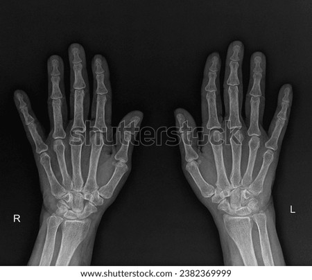
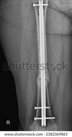
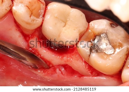
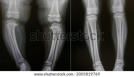
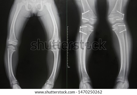
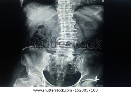
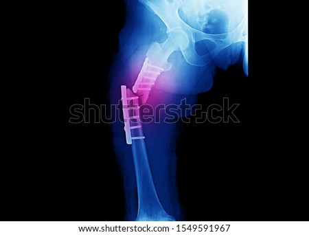
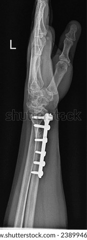
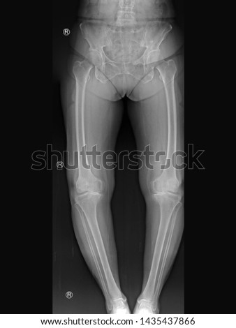
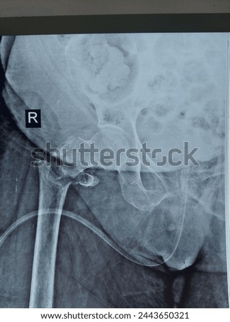
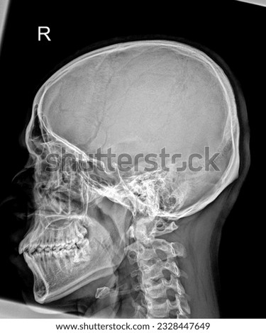
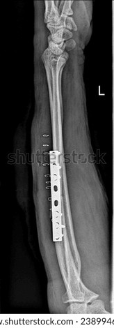
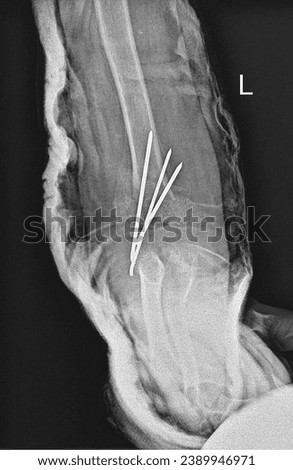
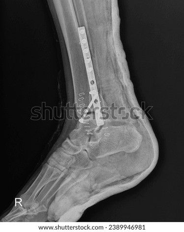
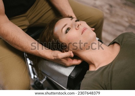
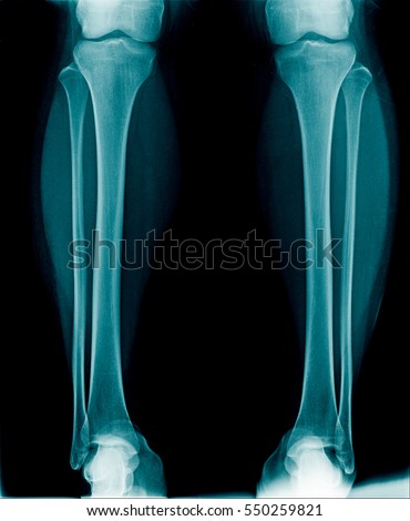
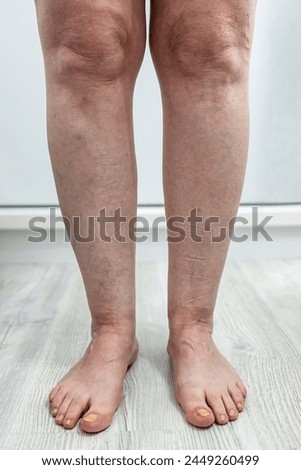
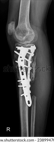
X-ray of the hand, demonstrating metacarpal and phalangeal bone alignment. Royalty-Free Stock PhotoMedical X-ray of a fractured femur bone, illustrating a common orthopedic injury. Royalty-Free Stock Photoconcept of Asian woman with Kyphosis: side view of laptop Work with hunched back, forward head posture, and spinal curvature Royalty-Free Stock PhotoOsteopath working on the back of a girl lying on an examination couch applying pressure to her bones realigning and manipulating them Royalty-Free Stock PhotoPeri-implantitis : Diseases that cause loss of bone around the dental implant, we can see the screw expose and gingival recession Royalty-Free Stock PhotoChiropractor doing adjustment on male patient Royalty-Free Stock PhotoChiropractor doing adjustment on female patient Royalty-Free Stock PhotoDental Dilemma: Missing and Damaged Teeth Royalty-Free Stock PhotoChiropractor doing adjustment on female patient Royalty-Free Stock PhotoConcept photo. Orthopedic pillow. Vertebral alignment. Improving the quality of sleep due to the use of the right rest products. Correct spine position Royalty-Free Stock PhotoThe x-ray show pre-operative and post-operative oblique osteotomy of proximal tibia of Blount's disease (LK II-III) in children. 3 years after surgey is good algnment and normal tibial plateau. Royalty-Free Stock PhotoThe x-ray of Rickets disease from hypophosphate is shown varus deformity and widening medial physis of bone. The other show improved alignment after the epiphysiodesis of both femurs and tibias. Royalty-Free Stock PhotoX-ray L-S spine Scoliosis and loss lordosis curve.Narrow L3-4-5 disc space with spur from degenerative change.Normal alignment.No fracture,bony destruction.No opaque calculi.Normal bowel gas pattern. Royalty-Free Stock PhotoHip and thigh x-ray showing closed fracture at shaft of right femur. The patient has underwent fixation with plate and screws. The image shows implant failure or broken plate at the fracture site. Royalty-Free Stock PhotoMan experiment terrible pain after breaking a molar Royalty-Free Stock PhotoOsteopathic therapist doing treatment to Caucasian woman with jaw problem, mandibular alignment. Treatment to relieve pain and improve the patient's health conditions. Royalty-Free Stock PhotoHigh-resolution X-ray capturing the successful fixation of a wrist joint fracture using a plate and screws. The image reveals the meticulous alignment of fractured wrist bones. Royalty-Free Stock Photofilm x-ray knee radiograph showing bow leg deformity (genu varus or bowlegged) from knee arthritis disease (osteoarthritis or OA disorder) which cause knee pain and walking problem.(R = right side) Royalty-Free Stock Photosubtrochanteric fracture of femur bone Royalty-Free Stock PhotoDetailed X-ray of the human skull showing intricate bone structure and dental alignment Royalty-Free Stock PhotoRadiographic view illustrating successful fixation of a forearm fracture using a plate and screws. The image highlights the precise alignment of the fractured forearm bones. Royalty-Free Stock PhotoDetailed X-ray illustrating successful fixation of an elbow joint fracture. The image showcases the precise alignment of the fractured elbow bones. Royalty-Free Stock PhotoOrthopedic X-ray capturing meticulous fixation of an ankle joint fracture using a plate and screws. The image showcases the precise alignment of the fractured ankle bones. Royalty-Free Stock PhotoChiropractor doing adjustment on female patient Royalty-Free Stock PhotoDoctor performs neck alignment on female patient in office Royalty-Free Stock Photox-ray both leg show normal alignment Royalty-Free Stock PhotoOsteopath doing a lumbosacral evaluation on a seated woman manipulating the muscles and spine of the lumbar and sacral regions of her lower back with his hands in close up Royalty-Free Stock PhotoUnderstanding Lower Limb Alignment,Anatomical Considerations for Health Care Professionals in Assessing Genu Varum and Bowed Feet Royalty-Free Stock PhotoLateral radiograph illustrating effective fixation of a proximal tibia and fibula fracture. The image reveals the precise alignment of the fractured bones, secured with orthopedic plate. Royalty-Free Stock PhotoMalocclusion, man in profile. Wrong bite: lower jaw extended forward and retracted. Bite correction with braces. Young man in profile Royalty-Free Stock Photo