Professional royalty-free CEREBELLUM stock photos and editorial news pictures from Shutterstock
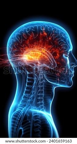
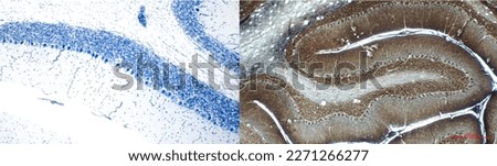
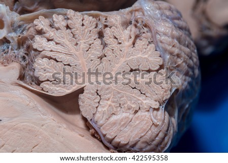
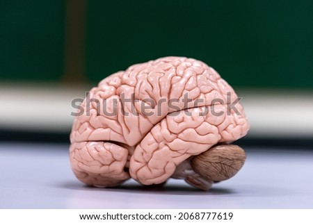
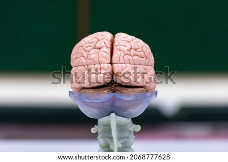
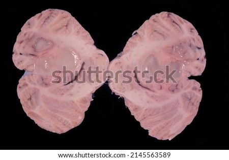
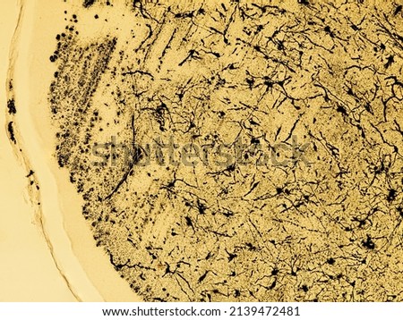
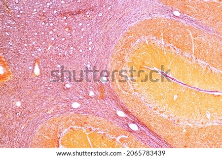
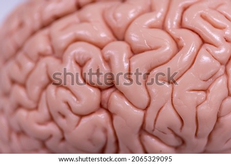
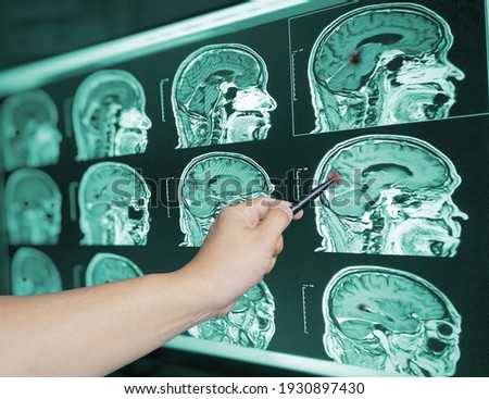
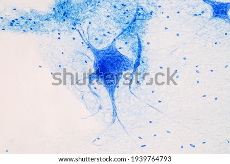
Neurologist hand pointing brain anatomy human model and brain disease lesion on white background.Part of human body model with organ system for health and doctor student study in university. Royalty-Free Stock Photo3D illustration of human brain on black background Royalty-Free Stock PhotoDoctor using pencil to demonstrate anatomy of human brain model in medical office. Human brain anatomical Model, behind view Royalty-Free Stock PhotoNeurologist hand pointing brain anatomy human model and brain disease lesion on white background.Part of human body model with organ system for health and doctor student study in university. Royalty-Free Stock PhotoPhoto of sagittal magnetic resonance imaging MRI of human brain level of corpus callosum with red highlight on cerebellum in patient with vertigo or dizziness. Blue tone dark background. Royalty-Free Stock PhotoCytoarchitectonics of the cerebellum. Histological sections are stained by the Nissl method (left) and by impregnation with silver nitrate (right). Royalty-Free Stock PhotoDoctor use robotic and innovative medical technology diagnose and examine patient brain with intelligence software. AI, Innovation medical healthcare and digital science technology in futuristic. Royalty-Free Stock PhotoClose-up detail of a sagittal section of the cerebellum Royalty-Free Stock PhotoCerebellum, Thalamus, Medulla oblongata, Spinal cord and Motor Neuron human under the microscope in Lab. Royalty-Free Stock PhotoCerebellum, Thalamus, Medulla oblongata, Spinal cord and Motor Neuron human under the microscope in Lab. Royalty-Free Stock PhotoHuman cerebellum showing two large cysts of liquefactive necrosis. In the brain, this type of necrosis can appear in bacterial infections or by the organization of an old cerebral infarction. Royalty-Free Stock Photo3D illustration of human brain on black background Royalty-Free Stock Photocat cerebellum cross section under the microscope - optical microscope x100 magnification Royalty-Free Stock PhotoLow magnification micrograph of a silver stained cerebellum. Each folium shows the three layers of cerebellar cortex (molecular, Purkinje and granular) surrounding a central axis of white matter. Royalty-Free Stock Photo
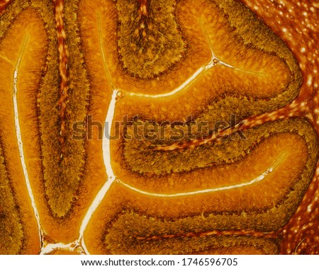
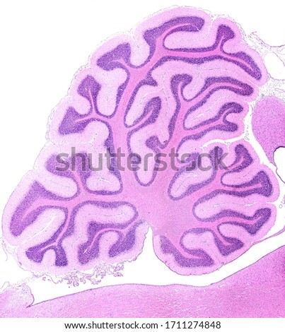
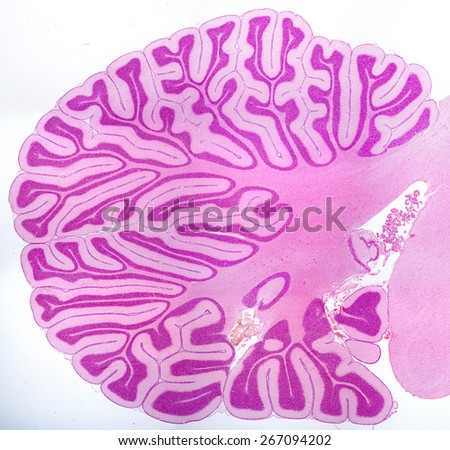
Hand touching brain and network connection on glitter bright lights colorful background Royalty-Free Stock PhotoCerebellum, Thalamus, Medulla oblongata, Spinal cord and Motor Neuron human under the microscope in Lab. Royalty-Free Stock PhotoCerebellum, Thalamus, Medulla oblongata, Spinal cord and Motor Neuron human under the microscope in Lab. Royalty-Free Stock PhotoClose up Hand doctor point brian scan image of a recent traumatic brain injury patient showing brain contusion and hemorrhage.Medical image concept. Royalty-Free Stock PhotoBrain Nervous System concept.Science is something that children should study and learn.Thinking process and Psychology of Kids. Royalty-Free Stock PhotoDoctor surgeon and neurologist checking brain testing result and medical diagnosis and examine patient brain with Science and technology, Digital medical healthcare. Royalty-Free Stock PhotoSagittal section of a rabbit cerebellum showing many ramified cerebellar folia. In each folium the molecular and granular layers and the central axis of white matter can be seen. HE stain Royalty-Free Stock PhotoEducation Spinal cord, Nerve, Cerebellum, Cortex and Motor Neuron Human under the microscope in Lab.
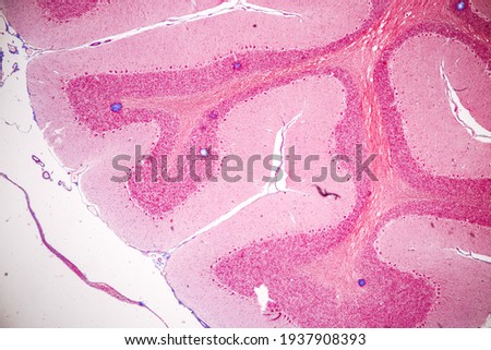
Royalty-Free Stock Photoartificial intelligence concept with a floating brain model Royalty-Free Stock PhotoNeurologist hand pointing brain anatomy human model and brain disease lesion on white background.Part of human body model with organ system for health student study in university.Medical education. Royalty-Free Stock PhotoAbstract palm hands touching brain with network connections, innovative technology in science and communication concept Royalty-Free Stock PhotoCerebellum sec, showing layers of cerebellum cortex, 1000x. Royalty-Free Stock PhotoSagittal section of a young cerebellum showing many ramified cerebellar lamellae. Light micrograph. H&E stain. Royalty-Free Stock PhotoEducation Spinal cord, Nerve, Cerebellum, Cortex and Motor Neuron Human under the microscope in Lab.
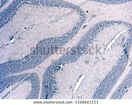
Royalty-Free Stock PhotoCross section of a cerebellum. Light micrograph. Cresyl Violet Staining (Nissl Staining). Royalty-Free Stock PhotoAbstract science. Networking, technology and innovation. Businessman holding brain in circle global network connection communication and data exchanges worldwide on modern interface background. Royalty-Free Stock Photo