Professional royalty-free CORNEAL stock photos and editorial news pictures from Shutterstock
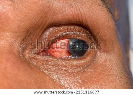
an optometrist uses a wide cone corneal topographer to take information from a patient's cornea. Royalty-Free Stock Photowoman eye with corneal dystrophy, keratoconus, thinning of the cornea. Royalty-Free Stock PhotoOptical biometer and full corneal topographer. measures axial length, corneal topography, pupillometry, corneal diameter and anterior chamber depth. Royalty-Free Stock PhotoCorneal topography Royalty-Free Stock PhotoA child undergoes a corneal topographer to take information about the cornea. Optometry. Royalty-Free Stock PhotoOphthalmology. Human eye close-up with red vascular mesh and inflammation on the eyeball. The concept of conjunctivitis and keratitis. Royalty-Free Stock Photoexamination of the elderly man on the corneal topographer. Videokeratograph is medical device for determining curvature of cornea. mandatory device for modern clinic or opticians shop. Royalty-Free Stock Photoclose up of the corneal ulcer during eye examination. Royalty-Free Stock PhotoA dark-skinned man is going to undergo a vision test using a corneal topograph to examine the cornea. Concept of corneal topographial in men. Royalty-Free Stock PhotoCorneal topographer shines red light for topography examination. Corneal topography eye vision test for visual description of the shape and power of the cornea Royalty-Free Stock Photowoman eye with corneal dystrophy, keratoconus, thinning of the cornea. Royalty-Free Stock PhotoCorneal ulcer by Herpes simplex Royalty-Free Stock PhotoMacro photo of the human eye with corneal disease keratoconus Royalty-Free Stock PhotoMacro eye photo. Keratoconus - eye disease, thinning of the cornea in the form of a cone. The cornea plastic. Royalty-Free Stock PhotoSelective Focus of Corneal Abrassion over Left Eye. Royalty-Free Stock PhotoMacro eye photo. Keratoconus - eye disease, thinning of the cornea in the form of a cone. The cornea plastic. Royalty-Free Stock PhotoClose up of Adorable Cavalier King Charles Spaniel with corneal ulcer eye injury looking up Royalty-Free Stock PhotoOptical biometer and full corneal topographer. measures axial length, corneal topography, pupillometry, corneal diameter and anterior chamber depth. Royalty-Free Stock PhotoCorneal moist chamber - special container for storage of human corneas. Corneas are collected from recently deceased individuals for transplantation (grafting) purposes. Royalty-Free Stock PhotoOptometrist is doing corneal topography. Corneal exam. Ophthalmology clinic. Background Royalty-Free Stock PhotoCorneal infection or ulcer called keratitis in Asian old woman. Royalty-Free Stock Photocloseup photo of the implantable collamer lens ICL for treating Royalty-Free Stock PhotoEye of a woman with cataract and corneal opacity Royalty-Free Stock PhotoCorneal topography also known as photokeratoscopy Royalty-Free Stock PhotoHuman eye of a woman affected by keratoconus, or conical cornea. Vision pathology leading to myopia and astigmatism and severe visual aberrations. Royalty-Free Stock Photoclose up of corneal abrasion during eye examination. in dark room. Royalty-Free Stock Photoan optometrist uses a wide cone corneal topographer to take information from a patient's cornea. Royalty-Free Stock PhotoMacro eye photo. Keratoconus - eye disease, thinning of the cornea in the form of a cone. The cornea plastic. Ophthalmology banner Royalty-Free Stock PhotoMacro eye photo. End stage of keratoconus. Keratoconus - eye disease, thinning of the cornea in the form of a cone. Royalty-Free Stock PhotoVitrectomy is a type of eye surgery to treat various problems with the retina and vitreous Royalty-Free Stock Photo