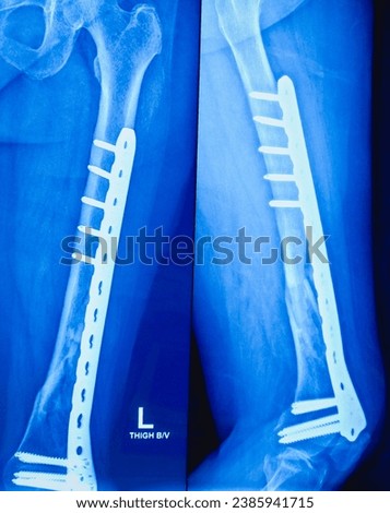
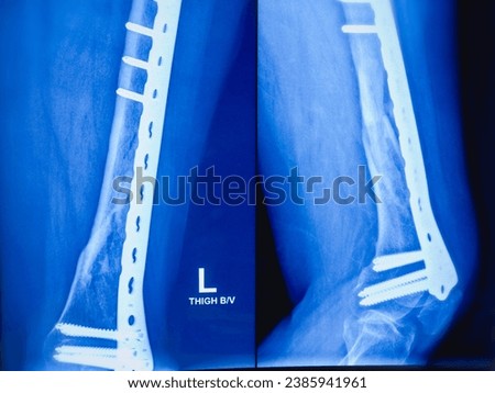
Professional royalty-free CORTICAL-BONE stock photos and editorial news pictures from Shutterstock
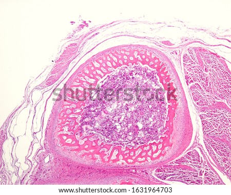
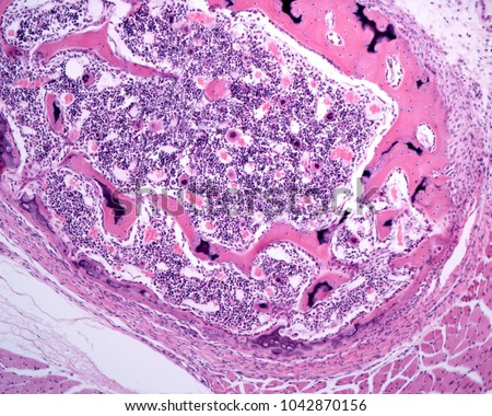
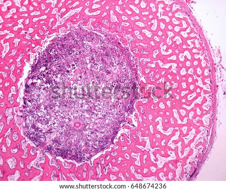
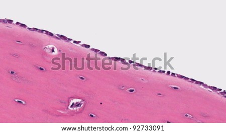
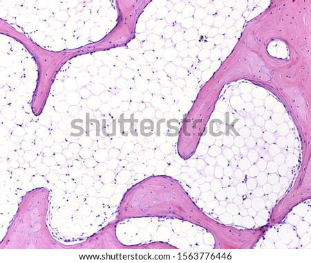
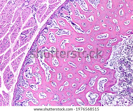
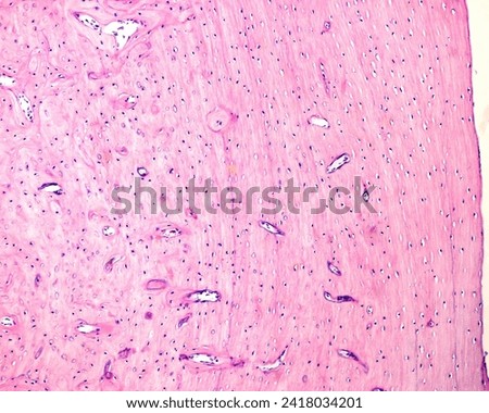
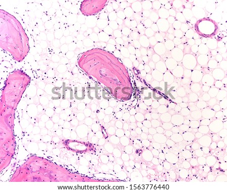
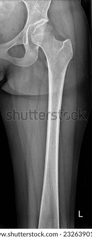
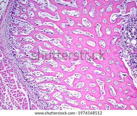
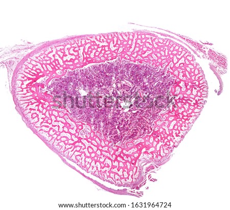
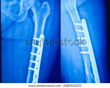
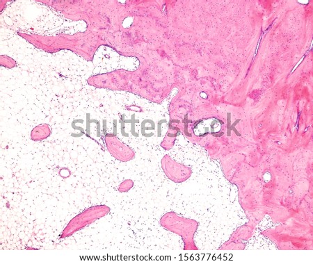
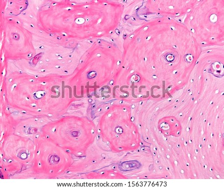
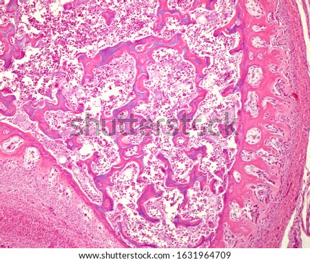
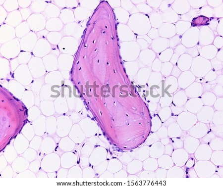
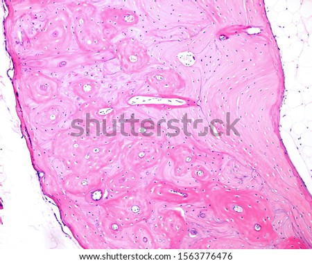
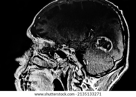
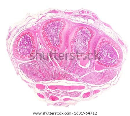
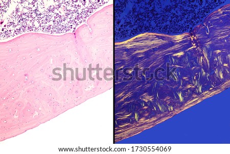
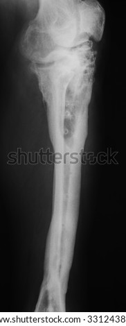
Low magnification micrograph showing a cross-sectioned immature bone. The periosteum surrounds the cortical of immature or primary woven bone tissue. The center material is the bone marrow. Royalty-Free Stock PhotoThe periosteum surrounds the cortical of immature or primary woven bone tissue (showing remains of cartilage). The intensely stained material of the center is the bone marrow. Royalty-Free Stock PhotoCross-section of the diaphysis of an immature bone. The periosteum surroundsthe cortical of immature or primary woven bone tissue. The intensely stained material of the center is the bone marrow. Royalty-Free Stock PhotoCompact cortical zone of a small bone. The round structures are osteons or Haversian systems, separated by interstitial lamellae. Parallel circumferential lamellae can be seen in the top left corner. Royalty-Free Stock PhotoBone forming cells osteoblasts lining the surface of mature cortical bone (pink substance in bottom half is bone). The bone has mature osteocytes in lacunae Royalty-Free Stock PhotoLight microscope micrograph showing a network of trabeculae of cancellous or spongy bone separated by large spaces occupied by adipose tissue of yellow bone marrow. Royalty-Free Stock PhotoIntramembranous ossification. Cross-sectioned immature bone (diaphysis). The periosteum surrounds the cortical of primary woven bone formed by a network of bone trabeculae. At right, the bone marrow Royalty-Free Stock PhotoBone. Near the surface of the compact bone, the bony lamellae are arranged parallel to the surface; these are called circumferential lamellae. Royalty-Free Stock PhotoTrabeculae of cancellous bone, also called trabecular or spongy bone separated by large spaces occupied by adipose tissue of yellow bone marrow. Royalty-Free Stock PhotoNormal Real X ray of Femur Royalty-Free Stock PhotoIntramembranous ossification. Cross-sectioned immature bone (diaphysis). The periosteum surrounds the cortical of primary woven bone formed by a network of bone trabeculae. At right, the bone marrow Royalty-Free Stock PhotoCross-sectioned immature bone (diaphysis). The periosteum surrounds the cortical of immature or primary woven bone tissue formed by a network of bone trabeculae. The center material is the bone marrow Royalty-Free Stock PhotoX-ray of Ankle view. Diffuse sclerosis with lytic area is noted in mid and distal shaft of the tibia results thickening of bony cortices with loss of medullary canal. Royalty-Free Stock PhotoX-ray of Ankle view. Diffuse sclerosis with lytic area is noted in mid, distal shaft of the tibia results thickening of bony cortices with loss of medullary canal. Soft tissue appears mildly swollen. Royalty-Free Stock PhotoX-ray of Ankle view. Diffuse sclerosis with lytic area is noted in mid, distal shaft of the tibia results thickening of bony cortices with loss of medullary canal. Royalty-Free Stock PhotoLimit between the cortical compact bone and the cancellous, trabecular or spongy bone. The spaces between bone trabeculae are occupied by adipose tissue of yellow bone marrow. Royalty-Free Stock PhotoCompact cortical zone of a small bone. The round structures are osteons or Haversian systems, separated by interstitial lamellae. Parallel circumferential lamellae can be seen in both surfaces Royalty-Free Stock PhotoComputed tomography (CT) of brain : SDH at left frontotemporoparietal convexity and right frontotemporal convexity / SAH in cortical sulci of left frontoparietotemporal lobe Royalty-Free Stock PhotoCAT SCAN with brain toxoplasmosis. Multiples brain tumors with ring enhancing lesion and perilesional edema. Located in mesencephalon and thalamus white matter, cortical and subcortical area. Royalty-Free Stock PhotoCortical of immature or primary woven bone tissue. In the center, still remain some mixed trabeculae showing a bluish center of calcified cartilage, covered by bone tissue stained with eosin. Royalty-Free Stock PhotoLight microscope micrograph showing a bone trabecula of cancellous or spongy bone tissue surrounded by yellow marrow. Bone lamellae and osteocytes can be seen in the trabeculae. Royalty-Free Stock PhotoCompact cortical zone of a small bone. The round structures are osteons or Haversian systems, separated by interstitial lamellae. Parallel circumferential lamellae can be seen in both surfaces Royalty-Free Stock PhotoCAT SCAN with brain toxoplasmosis. Multiples brain tumors with ring enhancing lesion and perilesional edema. Located in mesencephalon and thalamus white matter, cortical and subcortical area. Royalty-Free Stock PhotoVery low magnification micrograph showing a cross-sectioned palm of an embryo hand. The metacarpals show a cortical of immature or primary woven bone tissue, formed by a network of bone trabeculae Royalty-Free Stock PhotoComparative micrographs of cortical compact bone stained with HE (left) and seen under polarized light (right), which highlight the outer and inner circumferential lamellae and osteons Royalty-Free Stock PhotoComparative micrographs of cortical compact bone stained with HE (left) and seen under polarized light (right), which highlight the outer and inner circumferential lamellae and osteons Royalty-Free Stock PhotoCompact cortical zone of a small bone. The round structures are osteons or Haversian systems, separated by interstitial lamellae. Parallel circumferential lamellae can be seen in both surfaces Royalty-Free Stock PhotoCAT SCAN with brain toxoplasmosis. Multiples brain tumors with ring enhancing lesion and perilesional edema. Located in mesencephalon and thalamus white matter, cortical and subcortical area. Royalty-Free Stock PhotoCAT SCAN with brain toxoplasmosis. Multiples brain tumors with ring enhancing lesion and perilesional edema. Located in mesencephalon and thalamus white matter, cortical and subcortical area. Royalty-Free Stock PhotoX-ray image of knee include leg showing bone infection. Royalty-Free Stock Photo