Professional royalty-free ENDOPLASMIC stock photos and editorial news pictures from Shutterstock
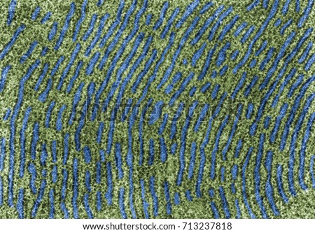
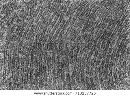
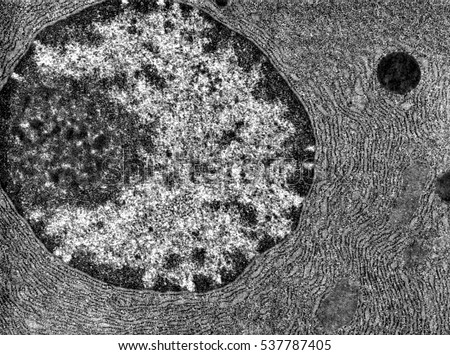
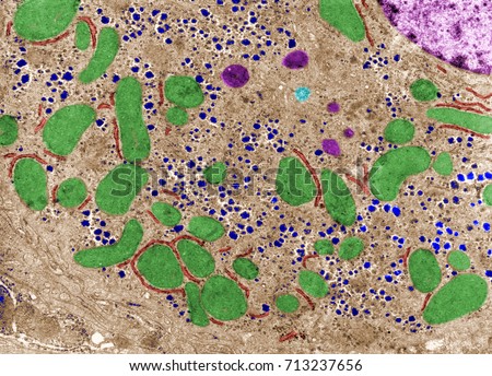
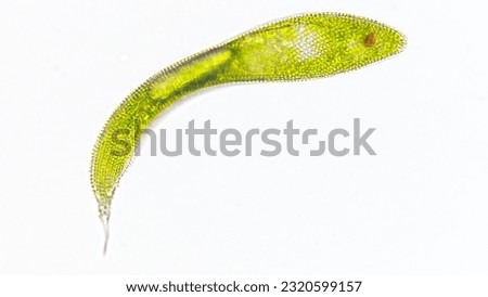
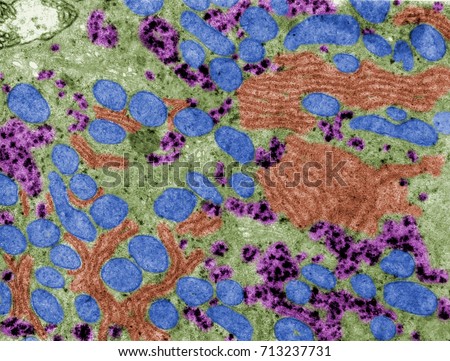
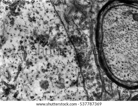
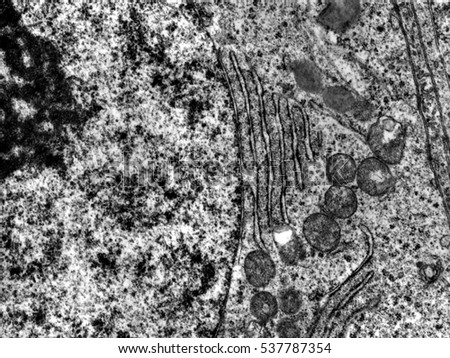
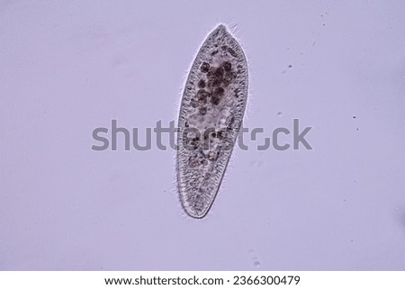
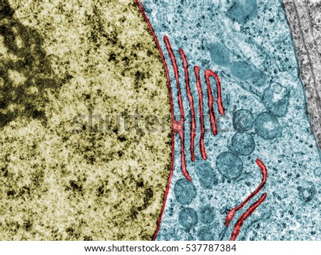
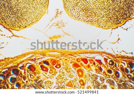
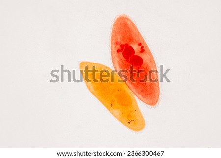
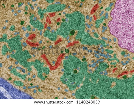
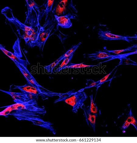
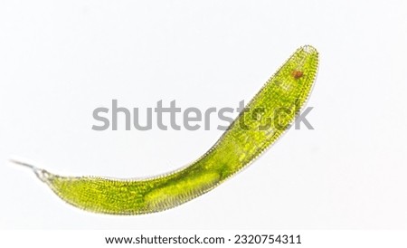
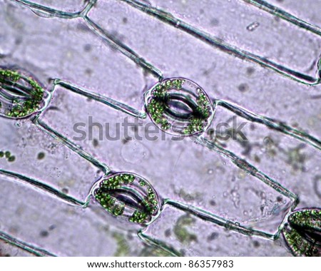
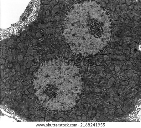
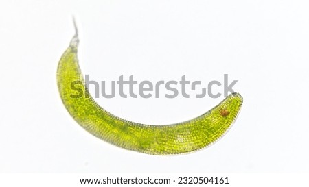
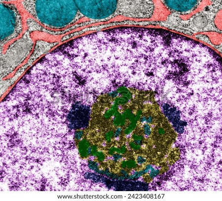
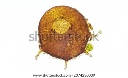
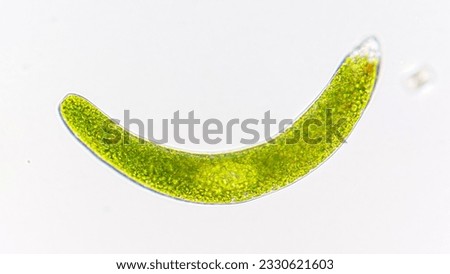
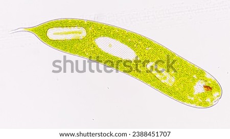
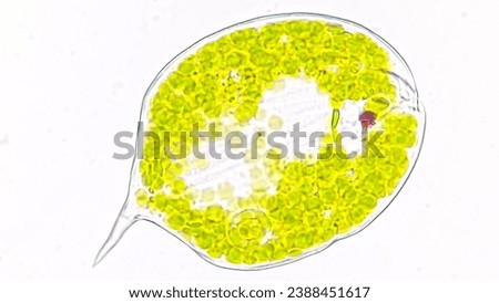
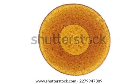
False colour TEM micrograph of a protein-synthetizing cell packed with parallel cisterns of RER with the lumen labelled in blue colour. The cytoplasm, full of ribosomes, is labelled in green color. Royalty-Free Stock PhotoTransmission electron microscope (TEM) micrograph showing the cytoplasm of a protein-synthetizing cell, full of parallel cisterns of rough endoplasmic reticulum. Royalty-Free Stock PhotoTEM showing the nucleus of a protein-synthesizing cell. The nuclear envelope, chromatin and nucleolus can be seen. The cytoplasm is full of RER. Royalty-Free Stock PhotoCell structure investigation: Microscopes allow visualizing microscopic cell structures such as nuclei, mitochondria, and endoplasmic reticulum. Royalty-Free Stock PhotoFalse colour transmission electron microscope (TEM) micrograph showing mitochondria (green), lysosomes (dark pink), glycogen (blue), rough endoplasmic reticulum (red) and a centriole (light blue). Royalty-Free Stock PhotoSingle cell flagellate eukaryotes, Euglena spirogyra. Live cell. Selective focus image Royalty-Free Stock PhotoFalse colour transmission electron microscope (TEM) micrograph showing mitochondria (blue), glycogen (pink), rough endoplasmic reticulum (red) in the cytoplasm of a hepatocyte. Royalty-Free Stock PhotoTransmission electron microscope (TEM) micrograph showing two synapses with clear synaptic vesicles. The postsynaptic element (a dendrite) shows ribosomes and cisternae of rough endoplasmic reticulum. Royalty-Free Stock PhotoTransmission electron microscope (TEM) micrograph showing a continuity between the nuclear envelope and a cistern of the rough endoplasmic reticulum. Royalty-Free Stock PhotoParamecium caudatum is a genus of unicellular ciliated protozoan and Bacterium under the microscope. Royalty-Free Stock PhotoFalse colour transmission electron microscope (TEM). Neuron cell body: nucleus (magenta), mitochondria (green), lysosomes (dark blue), multivesicular bodies (red), RER (brown) and Golgi (yellow). Royalty-Free Stock PhotoFalse colour transmission electron microscope micrograph showing a continuity between the nuclear envelope and a cistern of the rough endoplasmic reticulum (red). Nucleus (gold). Cytoplasm (blue). Royalty-Free Stock PhotoGolgi Apparatus. Microscopice- micrograph of a plant cell. Photo micro sections with high magnification with light microscope Royalty-Free Stock PhotoParamecium caudatum is a genus of unicellular ciliated protozoan and Bacterium under the microscope. Royalty-Free Stock PhotoFalse colour transmission electron microscope. Neuron cell body: nucleus (magenta), mitochondria (blue), lysosomes (dark green), RER (light green) and Golgi (red). Glial cell envelope (blue). Royalty-Free Stock PhotoImmunofluorescence confocal imaging of melanoma cancer cells Royalty-Free Stock PhotoSingle cell flagellate eukaryotes, Euglena spirogyra. Live cell. Selective focus image Royalty-Free Stock PhotoReal photo of plant cells and stoma with green chloroplast Royalty-Free Stock PhotoTEM micrograph showing a binucleated hepatocyte with many mitochondria and elongated cisterns of rough endoplasmic reticulum (RER). The space of Disse full of microvilli appear at three corners Royalty-Free Stock PhotoSingle cell flagellate eukaryotes, Euglena spirogyra. Live cell. Selective focus image Royalty-Free Stock PhotoProtein-secreting cell with nuclear envelope and RER (red), nucleolus fibrillar (green) and granular (yellow) and associated chromatin (dark blue), and secretory granules (light blue). Coloured TEM Royalty-Free Stock PhotoFreshwater testate amoeba, Centropyxis sp. Live cell. Stacked photo Royalty-Free Stock PhotoTestate amoeba species, Arcella sp. Live cell. Stacked image Royalty-Free Stock PhotoTestate amoeba, an amoeba with a smooth shell. Genus Centropyxis. Lugol fixed sample. 400x objective. selective focus Royalty-Free Stock PhotoTestate amoeba genus Difflugia. Stacked photo Royalty-Free Stock PhotoReal photo of plant cells and stoma.Pink plants cells. Royalty-Free Stock PhotoFreswater flagellate collected from pond water, Euglena ehrenbergii. 1040x magnification. Live cell. Selective focus Royalty-Free Stock PhotoFreshwater flagellates, Lepocinclis oxyuris (Basionym: Euglena oxyuris). Live cell. 1480x magnification. Selective focus image Royalty-Free Stock PhotoFreshwater flagellates, Phacus sp. Live cell. 2960x magnification. Selective focus image Royalty-Free Stock PhotoTestate amoeba species, Galeripora sp, an amoeba with a smooth shell. Live cell. 40x objective. Stacked image Royalty-Free Stock Photo