Professional royalty-free MULTINUCLEATED stock photos and editorial news pictures from Shutterstock
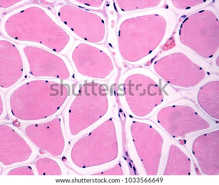
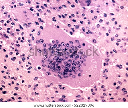
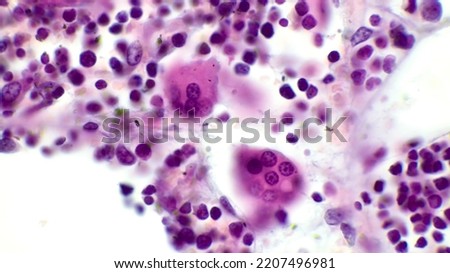
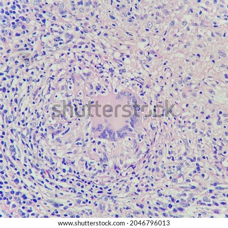
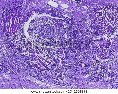
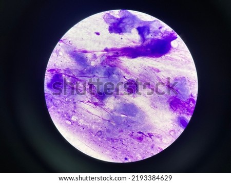
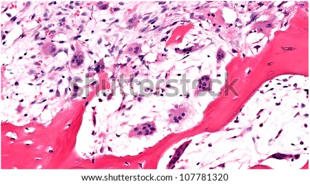
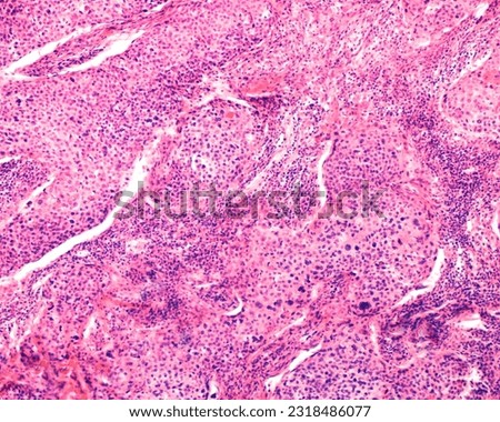
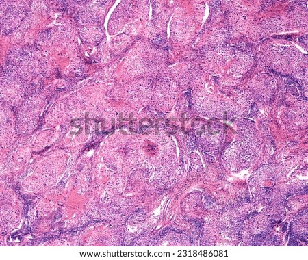
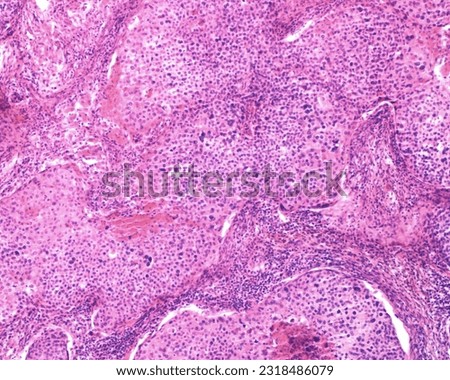
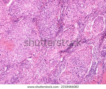
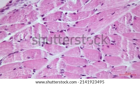
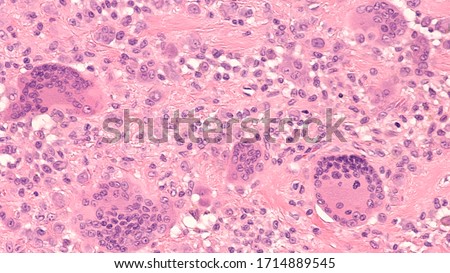
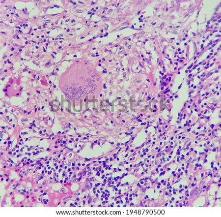
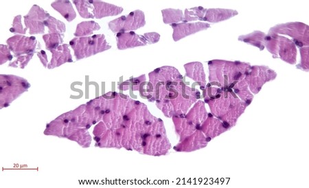
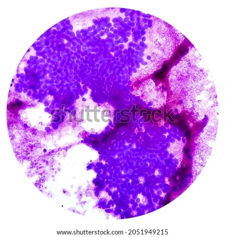
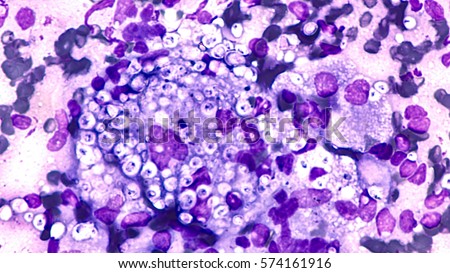
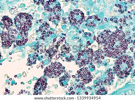
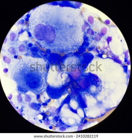
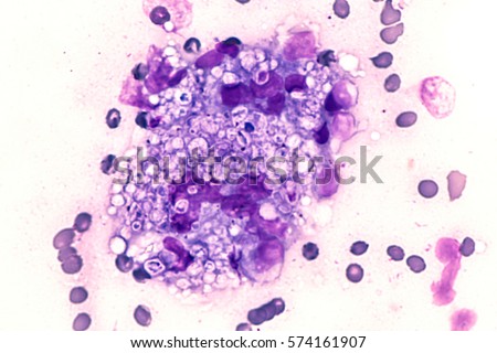
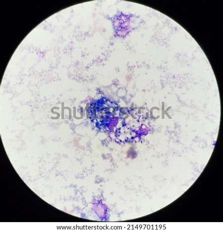
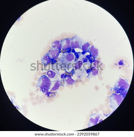
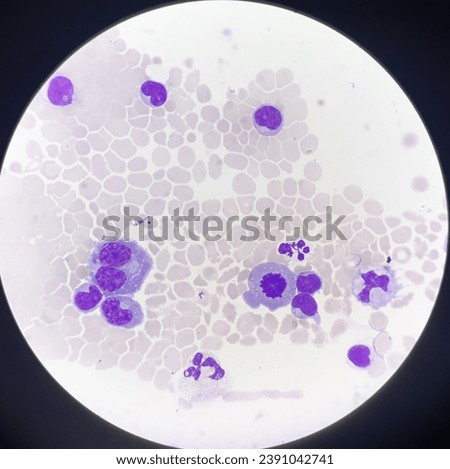
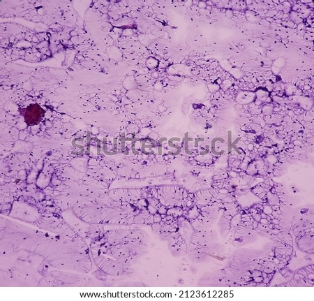
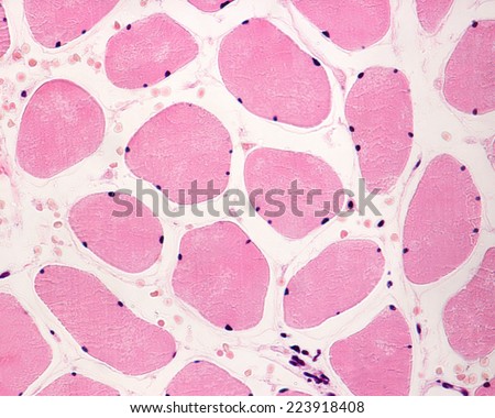
Amelanotic melanoma cytology from dog aspirate Royalty-Free Stock PhotoPanoramic web banner of microscope in medical research lab for science laboratory,study for making vaccine to protection a coronavirus COVID-19,scientist research in medical science laboratory concept Royalty-Free Stock PhotoSkeletal striated muscle fibers in cross section showing several peripheral nuclei (multinucleated cells). The endomysium, containing capillaries, appear as thin lines surrounding the muscle fibers. Royalty-Free Stock PhotoA mutinucleated giant cell derived from osteoclasts. Biopsy of a human giant cell tumour of bone. Light micrograph. Royalty-Free Stock PhotoOsteoclasts. Two osteoclasts in red bone marrow. The osteoclast is a large multinucleated cell which shows a ruffled border at a site of active bone resorption. H end E stain. Royalty-Free Stock PhotoCamera photo of multinucleated giant histiocyte in lymph node of patient with tuberculosis, magnification 400x, photograph through a microscope Royalty-Free Stock PhotoMultinucleated foreign body giant cells (FBGCs) in Hematoxylin and eosin staining tissues Royalty-Free Stock PhotoMultinucleated giant cells in Tzanck smear Royalty-Free Stock PhotoHighly magnified view of bone eating multinucleated osteoclasts along scalloped edges of trabecular bone due to osteoclastic bone resorption Royalty-Free Stock PhotoHuman ovary carcinoma. Light microscope micrograph of an ovarian malignant adenocarcinoma growing as solid cord masses. Tumor cells show nuclear atypia, giant nuclei and multinucleated cells. Royalty-Free Stock PhotoHuman ovary carcinoma. Light microscope micrograph of an ovarian malignant adenocarcinoma growing as solid cord masses. Tumor cells show nuclear atypia, giant nuclei and multinucleated cells. Royalty-Free Stock PhotoHuman ovary carcinoma. Light microscope micrograph of an ovarian malignant adenocarcinoma growing as solid cord masses. Tumor cells show nuclear atypia, giant nuclei and multinucleated cells. Royalty-Free Stock PhotoHuman ovary carcinoma. Light microscope micrograph of an ovarian malignant adenocarcinoma growing as solid cord masses. Tumor cells show nuclear atypia, giant nuclei and multinucleated cells. Royalty-Free Stock PhotoSkeletal striated muscle fibers in cross section showing the presence of several nuclei multinucleated cells located in the cell periphery. Light microscope photomicrograph. Royalty-Free Stock PhotoPhotomicrograph of a giant cell tumor of tendon sheath, with frequent multinucleated giant cells. Royalty-Free Stock PhotoPhoto of granuloma with multinucleated giant histiocytes, magnification 400x, photo under microscope Royalty-Free Stock PhotoSkeletal striated muscle fibers in cross section showing the presence of several nuclei multinucleated cells located in the cell periphery. Light microscope photomicrograph. Royalty-Free Stock PhotoHepatocellular dysplasia, cellular enlargement, nuclear pleomorphism, and multinucleation, light micrograph, photo under microscope Royalty-Free Stock PhotoFungal infection of lung, histoplasmosis: Fine needle aspirate (FNA) of a pulmonary nodule showing multinucleated giant cells containing small bubble-like organisms of Histoplasma capsulatum. Royalty-Free Stock PhotoLow magnification light microscope micrograph of cross sectioned skeletal muscle fibers showing the presence of many nuclei (multinucleated cells) located in the cell periphery. Royalty-Free Stock PhotoMicroscopic of disseminated histoplasmosis, a type of fungal infection by the fungus Histoplasma capsulatum. Yeast forms appear as black spherules within the cytoplasm of macrophages. GMS stain. Royalty-Free Stock PhotoAbnormal cells in body fluids. Royalty-Free Stock PhotoFungal infection of lung, histoplasmosis: Fine needle aspirate (FNA) of a pulmonary nodule showing multinucleated giant cells containing small bubble-like organisms of Histoplasma capsulatum. Royalty-Free Stock PhotoHepatocellular dysplasia, cellular enlargement, nuclear pleomorphism, and multinucleation, light micrograph, photo under microscope 100x view Royalty-Free Stock PhotoMacrophage cells in body fluid. Royalty-Free Stock PhotoAbnormal cells in body fluid. Royalty-Free Stock PhotoAbnormal cells in body fluids. Royalty-Free Stock PhotoAbnormal cells in body fluids. Royalty-Free Stock PhotoBreast lump(biopsy): Fat necrosis, injury of fatty tissue, show fragments of fatty tissue, foamy histiocytes, giant cells and inflammatory cells, benign condition. Royalty-Free Stock PhotoSkeletal striated muscle fibers in cross section showing the presence of several nuclei (multinucleated cells) located in the cell periphery. Light microscope photomicrograph Royalty-Free Stock Photo