Professional royalty-free VASCULAR-TISSUE stock photos and editorial news pictures from Shutterstock
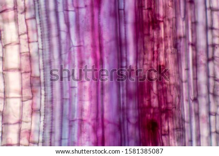
fern stem cross section under the microscope - optical microscope x200 magnification Royalty-Free Stock PhotoMonocot plant vascular tissue under the microscope view for education. Royalty-Free Stock PhotoPlant vascular tissue under the microscope view for education. Royalty-Free Stock Photo
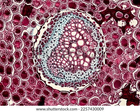
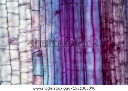
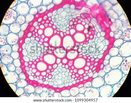
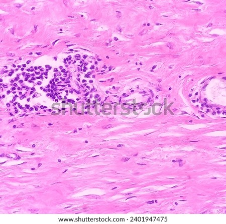
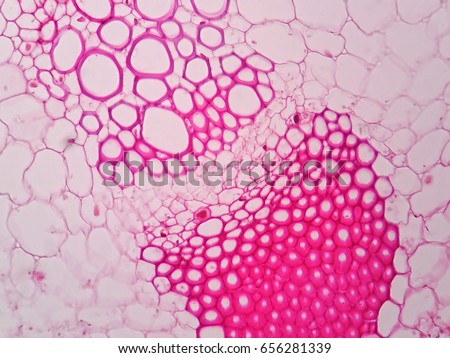
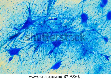
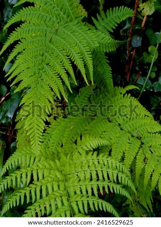
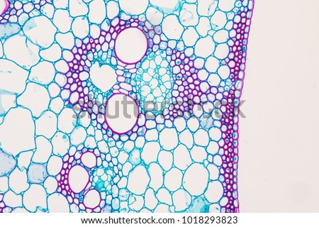
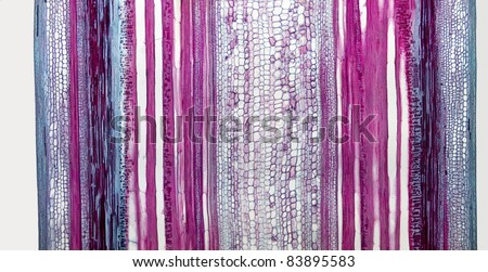
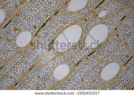
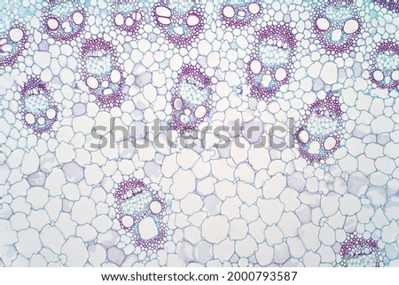
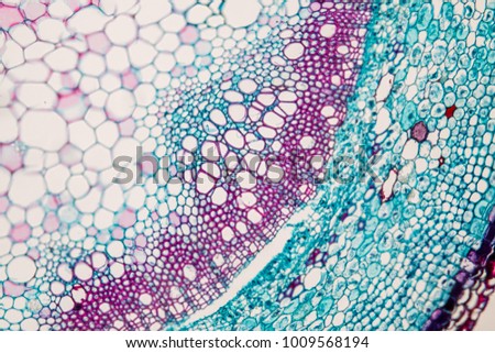
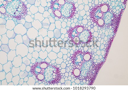
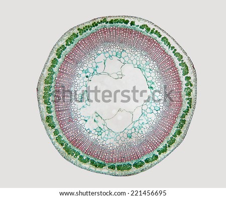
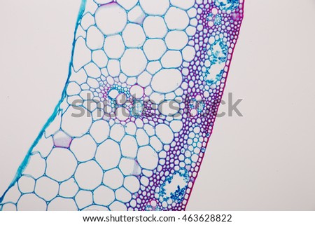
Cross-section Plant Stem under the microscope for classroom education. Royalty-Free Stock PhotoPlant vascular tissue under the microscope view for education. Royalty-Free Stock PhotoView in microscopy of vasular cylinder and cortex. section tissue of Ranunculus acris mature root. Royalty-Free Stock PhotoMicroscopic image of above knee amputation. Section show fibro collagenous tissue, fatty tissue, skeletal muscles with inflammatory cells. Bony soft tissue resection margin. Bone cancer. Royalty-Free Stock PhotoPlant vascular tissue under microscope view. Royalty-Free Stock PhotoScience background- neuron tissue. Nerve fibers: motor neurous- study with a large increase in the structural and functional nerve system. Core cell body processes. Scientific- spinal cord Royalty-Free Stock PhotoLongitudinal section through cells of a stem from a maize plant (Zea mays) under the microscope. Royalty-Free Stock PhotoPhoto ferns, are vascular plants members of the pteridophyte taxon. They have vascular tissues, true leaves, reproduce through spores and do not produce seeds or flowers. Royalty-Free Stock PhotoCross-section Plant Stem typical Monocot and Dicot under the microscope for classroom education. Royalty-Free Stock PhotoA detailed longitudinal section of cinnamon bark (Cinnamomum sp) showing associated structures and layers. Magnification 10x Royalty-Free Stock Photophoto of a plant tissue under the microscope
Royalty-Free Stock PhotoMonocot plants stem show plant vascular tissue under light microscope view for botany education. Royalty-Free Stock Photo
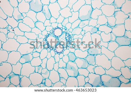
Cross-section Plant Stem under the microscope for classroom education. Royalty-Free Stock PhotoParenchyma Tissue of plant under the microscope Royalty-Free Stock Photo
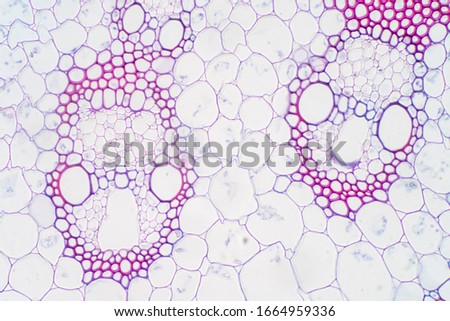
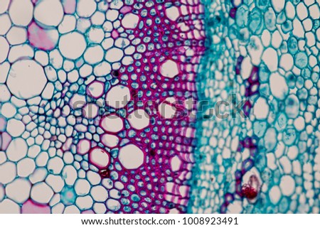
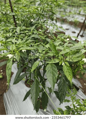
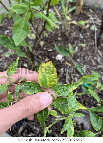
Cross-section Plant Stem under the microscope for classroom education. Royalty-Free Stock PhotoCross-section Plant Stem typical Monocot and Dicot under the microscope for classroom education. Royalty-Free Stock PhotoBroken capillaries on the face, Red skin problems, Congested skin. The concept of health problems, spider veins, macro, close up Royalty-Free Stock PhotoFerns, or ferns, are vascular plants that are members of the pteridophyte taxon (which no longer have taxonomic validity and are only used as an informal name). They have vascular tissues (xylem and p Royalty-Free Stock PhotoFusarium wilt is a disease that the fungus assaults the vascular tissue within the root discoloration. Royalty-Free Stock Photoa chili pepper plant stem is usually cylindrical, green, and covered with small ridges. It serves as the main support for the plant's leaves, flowers, and fruits. The stem contains vascular tissues th Royalty-Free Stock PhotoInfected plants may be stunted and will begin to wilt because the plant is unable to uptake water. Vascular tissue may show discoloration (i.e., browning). Royalty-Free Stock PhotoThis picture is a cross section of stem plant tissue and under a light microscope.
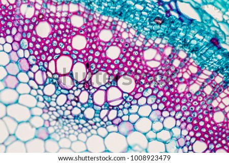
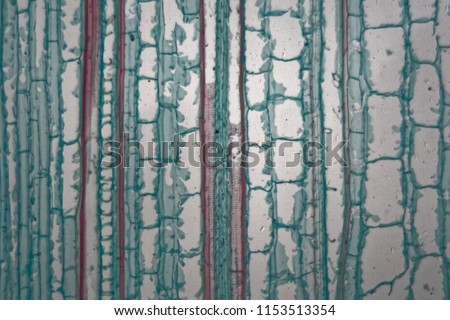
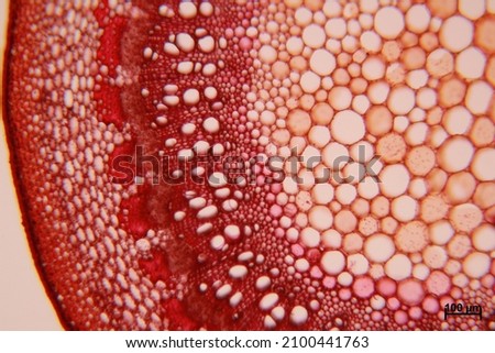
Found in many types of plant tissues, such as ground tissue (parenchyma cell, collenchyma cell, sclerenchyma) Royalty-Free Stock PhotoDoctor makes ultrasound of wrist in clinic closeup Royalty-Free Stock PhotoThe common Flax ( Linum usitatissimum) stem, cross section. Magnification 40x. Royalty-Free Stock PhotoAbstract micrograph with polarization of a bract from a Protea flower, showing the midrib vascular tissue (orange), like magma in a leaf, at 40x. Royalty-Free Stock PhotoAbstract micrograph with polarization of a bract from a Protea flower, showing vascular tissue at 100x. Royalty-Free Stock PhotoSclerenchyma Tissue under the microscope Royalty-Free Stock Photo