Professional royalty-free RECEPTOR stock vectors and illustrations from Shutterstock
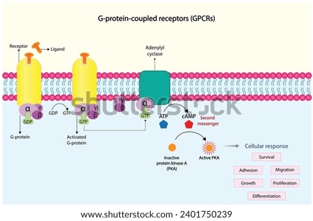
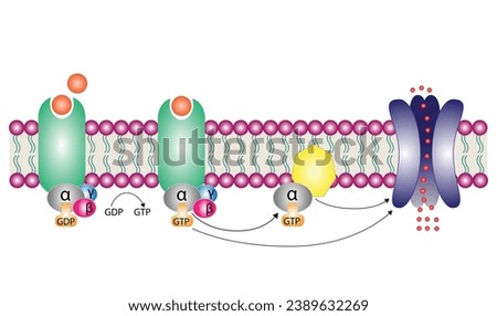
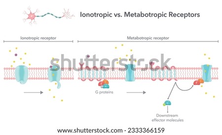
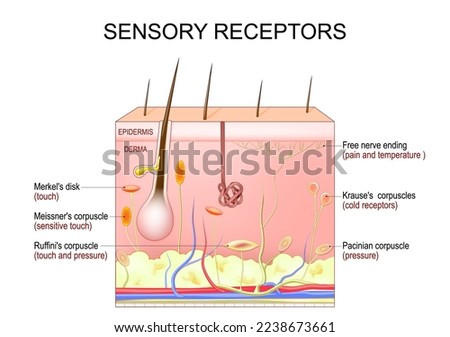
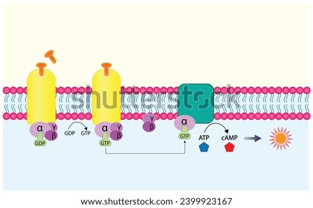
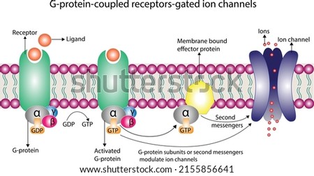
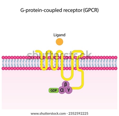
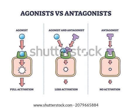
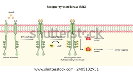
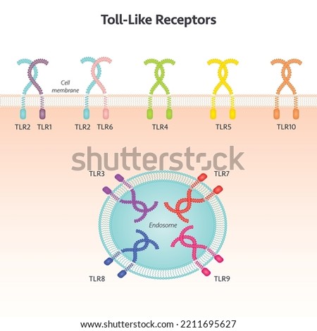
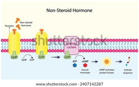
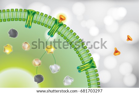
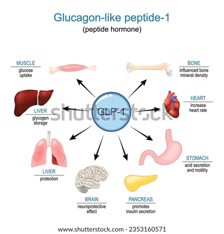
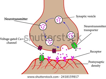
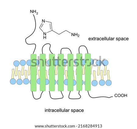
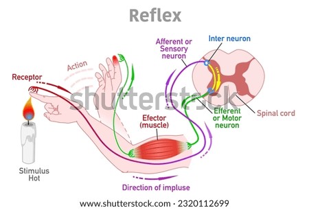
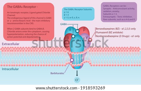
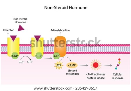
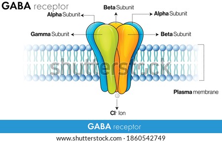
G protein coupled receptors gated ion channel. Structure of a G protein-coupled receptor (GPCR). Mechanism for the transport of ions. Cell membrane receptors for ligands bind. vector illustration. Royalty-Free Stock PhotoG protein coupled receptors gated ion channel. Structure of a G protein-coupled receptor (GPCR). Mechanism for the transport of ions. Cell membrane receptors for ligands bind. vector illustration Royalty-Free Stock PhotoVector line icon for receptor Royalty-Free Stock PhotoIonotropic versus metabotropic receptors vector illustration diagram Royalty-Free Stock Photoskin sensory receptors. Cross section of humans skin layers with Free nerve ending, Merkel's disk, Pacinian, Ruffini's, Krause's, and Meissner's corpuscles. Vector illustration Royalty-Free Stock PhotoG protein coupled receptor. Structure of a G protein-coupled receptor (GPCR). Cell membrane receptors for ligands binding. cAMP, second messenger, production amplification. vector illustration. Royalty-Free Stock PhotoG protein coupled receptors gated ion channel. Structure of a G protein-coupled receptor (GPCR). Mechanism for the transport of ions. Cell membrane receptors for ligands bind. vector illustration Royalty-Free Stock PhotoG protein coupled receptors gated ion channel. Structure of a G protein-coupled receptor (GPCR). Mechanism for the transport of ions. Cell membrane receptors for ligands bind. vector illustration Royalty-Free Stock PhotoSynapse Icon. Thin Linear Illustration for Neurological Connections, Brain Function, and Neural Communication Education. Isolated Outline Vector Sign. Royalty-Free Stock PhotoAgonists vs antagonists drugs behavior to receptor activation outline diagram. Labeled educational pharmacological compounds effect to block or stimulate body vector illustration. Pain cure or relief. Royalty-Free Stock PhotoTyrosine kinase receptor. Dimerization, phosphorylation, activation and cellular response. Cell membrane receptors for ligands as growth factors and cytokines binding. Insulin receptor. vector design Royalty-Free Stock PhotoToll-like receptor science vector illustration background graphic Royalty-Free Stock Photoendocytosis: Cells that undergo receptormediated endocytosis have pits coated with the protein clathrin that initiate endocytosis when target molecules bind to receptor proteins. Royalty-Free Stock PhotoG protein coupled receptor (GPCR). Cell membrane receptors for ligands binding. cAMP, second messenger, production amplification. Protein kinase A, PKA. cAMP response element binding protein (CREB). Royalty-Free Stock PhotoReceptor cells. Sense organs examples. Vision, various nerve cells. Touch, meissner corpuscles include, rods, cones, olfactory smell, hair, hearing, gustatory, taste. Colored illustration vector Royalty-Free Stock PhotoT-cell and Chimeric antigen receptor T cell ,CAR T cell therapy, for use in immunotherapy. chemotherapy. vector illustration. Royalty-Free Stock PhotoNonsteroid hormones mechanism of action. The hormone is the first messenger, binds to the receptor and activating a second messenger inside the cell resulting in cellular response. Vector illustration Royalty-Free Stock Photoantibody blocking cell receptor Royalty-Free Stock PhotoGLP-1. Glucagon-like peptide-1 for Appetite regulation, weight loss and Treatment of diabetes. peptide hormone Functions and effects on Human internal organs. vector diagram Royalty-Free Stock PhotoStructure of a typical chemical synapse.Vector illustration. Royalty-Free Stock PhotoOlfactory nerves with sensory facial nose organs anatomy outline diagram. Labeled educational scheme with human head nasal scent system and plane, cavity or bulb medical location vector illustration. Royalty-Free Stock PhotoSchematic structure of histamine receptor which belongs to GPCR receptors. Vector illustration. Royalty-Free Stock PhotoNeurotransmitters are released from synaptic vesicles of the presynaptic neuron and bind to receptors on the postsynaptic neuron, triggering an impulse through the 2nd neuron. Royalty-Free Stock PhotoReceptor cells: rod and cone (Vision), Meissner's corpuscle (touch), Olfactory receptor (smell), hair cell (Hearing) and gustatory cell (taste). Human anatomy Royalty-Free Stock PhotoReflex arc, action. Somatic receptors in the skin, muscles and tendons, message to brain, pathway. Stimulus, hot, touch. sense, effector muscle, spinal cord, sensory motor neuron. Illustration vector Royalty-Free Stock PhotoGABA A Receptor Diagram in cell membrane with explanation about binding sites, ligands and activity. Infographic pharmacology, science, health care vector illustration of neurotransmitter in the CNS. Royalty-Free Stock PhotoNonsteroid hormones mechanism of action. The hormone is the first messenger, binds to the receptor and activating a second messenger inside the cell resulting in cellular response. Vector illustration Royalty-Free Stock PhotoWebsite template. Human nerve translucent low poly triangles. Futuristic glowing organ hologram on dark blue background. Medical innovation diagnosis treatment concept. Banner vector. Royalty-Free Stock PhotoHuman nerve cell red translucent low poly triangles on dark blue background. Futuristic glowing organ hologram and copy space for text. Medical and science concept. Banner design vector. Royalty-Free Stock PhotoGABA receptor Ultra Structure vector design Royalty-Free Stock Photo